The Muscle–Brain Message: What Irisin Research Actually Shows
An exercise-linked signal produced intriguing results in Alzheimer’s mouse models. Here is what the experiments establish—and what they cannot tell us about treating people.
A mouse remembering an unfamiliar object, a sample of cerebrospinal fluid, and an exercise-linked protein: the irisin story brings together measurements that answer different questions. Treating them as one continuous proof of a human treatment makes the story sound simpler than the research actually is.
Irisin is a signaling protein produced from a precursor called FNDC5. A 2019 Nature Medicine paper investigated its relationship to synaptic function and memory in experimental Alzheimer’s disease models, alongside observations in human samples. The useful question is what these experiments establish, not whether a biological mechanism makes an appealing headline. [1]
What the researchers actually tested
The experimental work included lowering and increasing FNDC5/irisin signaling in mice. Reducing the pathway impaired measures of synaptic plasticity and object-recognition memory. Increasing it rescued memory and synaptic deficits in Alzheimer’s mouse models. Blocking it also weakened the beneficial effects of exercise in those models. These manipulations support a role for the pathway within the experimental systems tested. [1]
The human findings answered a different question. FNDC5/irisin was reduced in Alzheimer’s hippocampal tissue and cerebrospinal fluid. That comparison does not establish that low irisin initiated the disease, or that raising it would improve a person’s symptoms. The study did not administer an irisin treatment to patients and compare their clinical outcomes with a control group. [1]
Mechanism atlas: structure, expression and signaling
The following figures separate three types of evidence. A measured protein configuration tells us about physical structure. Gene-expression experiments show changes in the amount of RNA produced. Signaling experiments measure activity or chemical modification of existing proteins. An upward arrow does not mean that every part of the pathway increased, and a phosphorylation change does not necessarily mean that more protein was made.
1. What irisin actually looks like
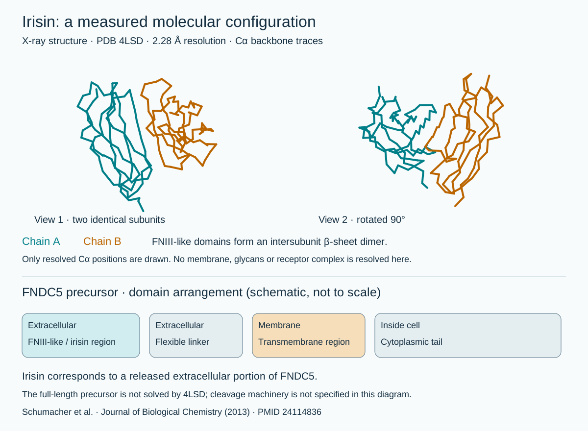
The crystallographic study resolved an FNIII-like fold and a two-subunit interface involving a shared β-sheet. Our illustration plots measured backbone coordinates from PDB 4LSD, rather than an AI-invented molecular shape. The membrane precursor below is a separate domain schematic. The structure does not show a brain receptor bound to irisin. [2: Journal of Biological Chemistry, 2013]
2. Upregulation, downregulation and a possible feedback loop
In the 2013 experiments, endurance exercise increased hippocampal expression of PGC-1α and FNDC5 in mice. PGC-1α is a transcriptional coactivator: it works with transcription factors, including ERRα, rather than binding DNA on its own. Increasing neuronal FNDC5 raised Bdnf expression; reducing FNDC5 with RNA interference lowered it. These arrows summarize experimental regulation, not direct binding between the labeled molecules. [3: Cell Metabolism, 2013]
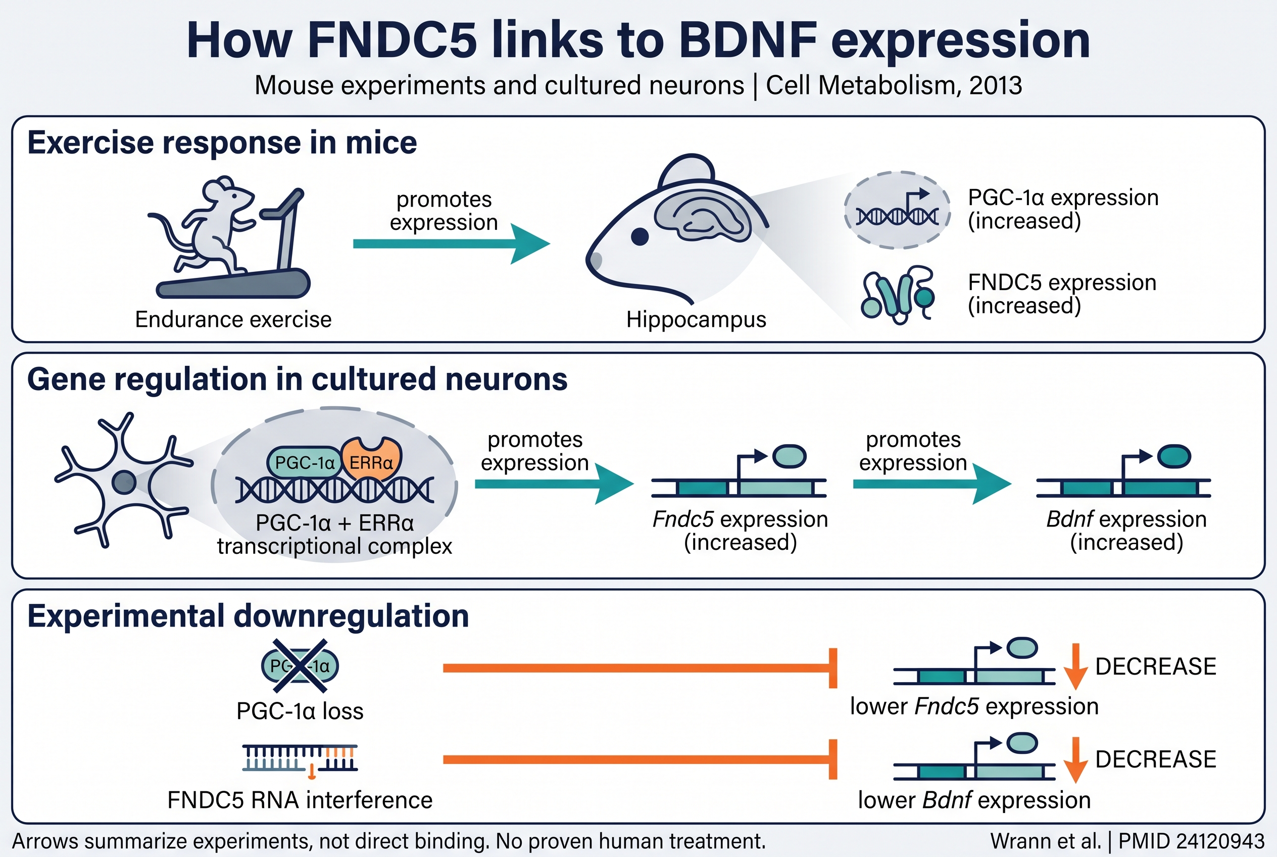
The same study found that recombinant BDNF lowered Fndc5 expression in cultured cortical neurons. This supports a possible negative-feedback relationship: FNDC5 promotes Bdnf expression, while BDNF exposure can reduce Fndc5 expression. It does not establish a universal feedback strength, a human dose response or a reason to maximize either signal. [3]
3. Activation and suppression are different measurements
The 2019 paper reported increased cAMP, PKA activity and CREB phosphorylation after recombinant irisin exposure in human cortical slices maintained outside the body. In mouse hippocampal slices, PKA inhibition blocked the CREB phosphorylation response. Those results support signaling involvement; tissue taken from people is not the same as treating people. [1: Nature Medicine, 2019]
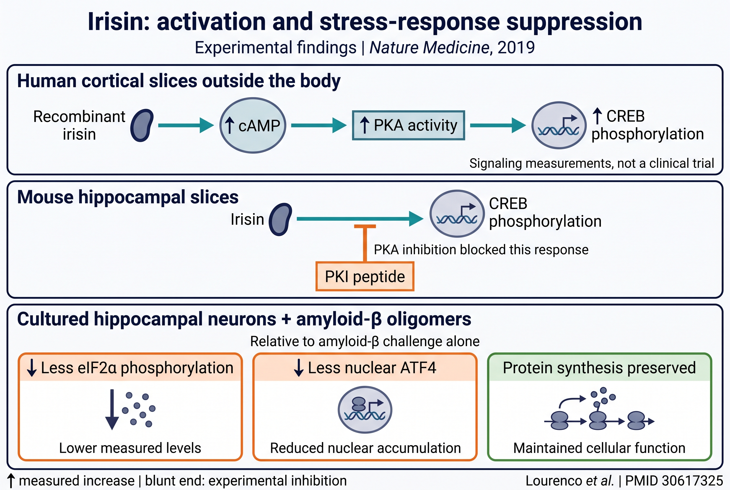
The paper did not identify the cellular receptor responsible for these brain responses. We therefore do not insert a particular receptor, a receptor docking pose or an uninterrupted muscle-to-human-memory chain into this figure. The pathway maps are interpretations of the cited experiments, not measurements of every intervening step.
Figure production: the molecular backbone is rendered from experimental PDB coordinates. The two pathway illustrations were created with Nano Banana Pro and checked against the primary papers. They use conceptual shapes and should not be interpreted as molecular imaging or clinical results.
Why the distinction matters
An experimental manipulation can be persuasive evidence for a mechanism without being a validated treatment. In a mouse experiment, researchers can control exposures and examine processes that would be difficult to isolate in people. Those strengths help answer a mechanistic question. They do not remove the next question: whether the same intervention, delivered safely and feasibly to a person, produces a worthwhile clinical benefit.
Likewise, finding a difference in a biological sample is a starting point for explanation. The difference could be part of the disease process, a consequence of it, or connected to other changes. A treatment claim needs evidence about what happens when researchers deliberately intervene, not simply a comparison between samples from different groups.
A memory task is not the whole clinical outcome
“Memory improved” needs an object: improved on which test, in which species, and compared with what? Object-recognition behavior is an experimental outcome. Daily functioning, quality of life, symptom progression and adverse effects are different outcomes. A useful account of this paper names its tests rather than replacing them with a broader promise of restored human cognition.
Our interpretation is that the most interesting next step is not a stronger slogan. It is an experiment that connects the mechanism to a patient-relevant result. Researchers would need to establish a practical intervention, its safety, an appropriate comparison group and meaningful outcomes over sufficient follow-up. This is a framework for assessing future evidence, not a claim that those requirements have already been met.
Does this tell you which workout is best?
No. This paper cannot rank everyday leg workouts for preventing dementia in people. To establish that one routine is superior, researchers would need to compare the routines in a relevant human population and measure the outcome being claimed. A pathway associated with exercise cannot supply that missing head-to-head comparison.
The same reasoning applies to products marketed around a mechanism. Naming irisin on a sales page would not demonstrate that a product safely changes the pathway, reaches the relevant tissue or improves health. This article makes no recommendation to buy or self-administer an irisin product.
Watch the short explanation
The video uses synthetic narration and Nano Banana Pro conceptual imagery. Its glass brain is an illustration, not a scan or an experimental result. The article contains the fuller explanation and sources.
How to read the next irisin headline
Start with the population: cells, animals, human samples or treated patients. Then identify the intervention and comparator, followed by the measured outcome. Finally, separate what the authors observed from what the headline predicts. These steps let a promising mechanism remain promising without turning an unanswered clinical question into an established remedy.
The mouse task and the human sample both matter. Their value comes from keeping their answers distinct.
By the Magellan Longevity Editorial Desk. AI-assisted original research explainer. This is a focused reading of three primary papers, not a systematic review of exercise or dementia treatments. No product recommendation or researcher endorsement.
Educational, not medical advice.
The takeaway
Irisin is a research lead supported by mechanistic experiments, not a proven dementia treatment or a basis for naming a best leg workout.
References
3 peer-reviewed sources, published 2013–2019, across 3 journals. Every citation links to its PubMed record.
- Exercise-linked FNDC5/irisin rescues synaptic plasticity and memory defects in Alzheimer's models. ↗Nature Medicine · 2019 · PMID 30617325 · DOI 10.1038/s41591-018-0275-4
- Journal of Biological Chemistry · 2013 · PMID 24114836 · DOI 10.1074/jbc.M113.516641
- Cell Metabolism · 2013 · PMID 24120943 · DOI 10.1016/j.cmet.2013.09.008
Mechanisms and molecules in this article
Each links to its Magellan monograph — what it is, what it does, and the studies behind it.
Related reading
The most widely cited number in the caregiving literature, a 63 percent rise in mortality, belonged to…
Rentosertib has renewed the debate over biological age. A randomized human experiment offers an important…
Tea, berries and spices were associated with a changing DNA-methylation clock in a small lifestyle trial.…
Prefer the interactive version? Open this article inside the Magellan app →