PTCHD4 and Aging: A Promising Target With a Complicated Backstory
A new study connects a membrane protein to AKT signaling, senescence and mouse lung fibrosis. Earlier human-cell experiments show why the direction of intervention still needs careful testing.
A membrane protein. A signaling enzyme. A damaged lung. The new PTCHD4 paper places these three pieces in the same aging story, but the most revealing part of that story may be what happens when a different laboratory studies the same protein in a different kind of cell.
In Aging Cell, Wang and colleagues report that lowering PTCHD4 reduced senescence-related changes in their experimental models. A 2024 paper in Nucleic Acids Research found that silencing it increased growth arrest and DNA damage in pre-senescent human fibroblasts. These results make PTCHD4 an interesting research target. They also make a universal “turn it down to slow aging” interpretation premature.
The evidence boundary: this is a preclinical research update, not a treatment recommendation. For the new paper, this article uses the independently verified PubMed abstract; the publisher’s full text was not accessible during preparation. The earlier paper was read in full through PubMed Central. Sample sizes, intervention schedules, detailed survival analyses and effect sizes from the new paper remain unverified here.
What the new study actually reports
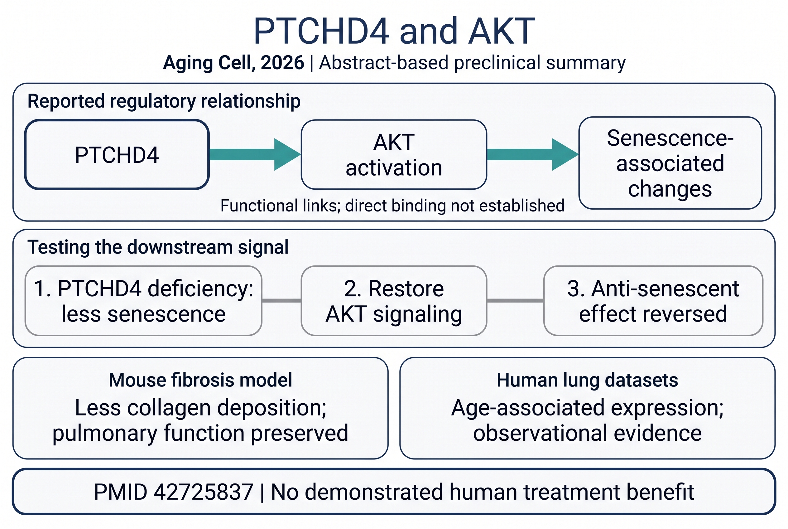
PTCHD4 expression increased in several cellular senescence models and with age in mouse tissues and human lung gene-expression datasets. Experimentally reducing the protein’s function attenuated senescence progression, while increasing its expression promoted it. The distinction matters: expression patterns describe an association; changing a candidate regulator tests whether it can influence the process. Aging Cell, 2026.
The investigators also report benefits of PTCHD4 deficiency in D-galactose-induced aging-related mouse phenotypes and a bleomycin-induced lung fibrosis model. The latter included less collagen deposition and preservation of pulmonary function. The abstract additionally reports an extension of median mouse lifespan, but does not provide the information needed here to evaluate the magnitude or survival-study design. We therefore give no lifespan percentage and do not label this a demonstration of extended healthy human life.
For a reader, the important separation is between an experimentally induced condition and ordinary aging. A result under a defined laboratory challenge can identify a useful mechanism without establishing how a proposed intervention will work across tissues, ages or diseases. Human lung datasets add relevance; they do not supply a trial in which people received a PTCHD4-targeted treatment.
The strongest mechanistic clue is a rescue experiment
The new abstract links PTCHD4 to increased activity of AKT, a signaling protein. Restoring AKT signaling reversed the anti-senescent effects of PTCHD4 deficiency. This is more informative than simply finding two markers elevated together: the investigators changed a downstream signal and tested whether the phenotype changed with it.
Our interpretation is that this supports AKT as a functional participant in the tested system. It does not establish that PTCHD4 physically binds AKT, identify a drug-binding pocket, or prove that this pathway is the sole explanation for every reported outcome. Those are separate claims requiring separate experiments.
The earlier paper makes the story less simple
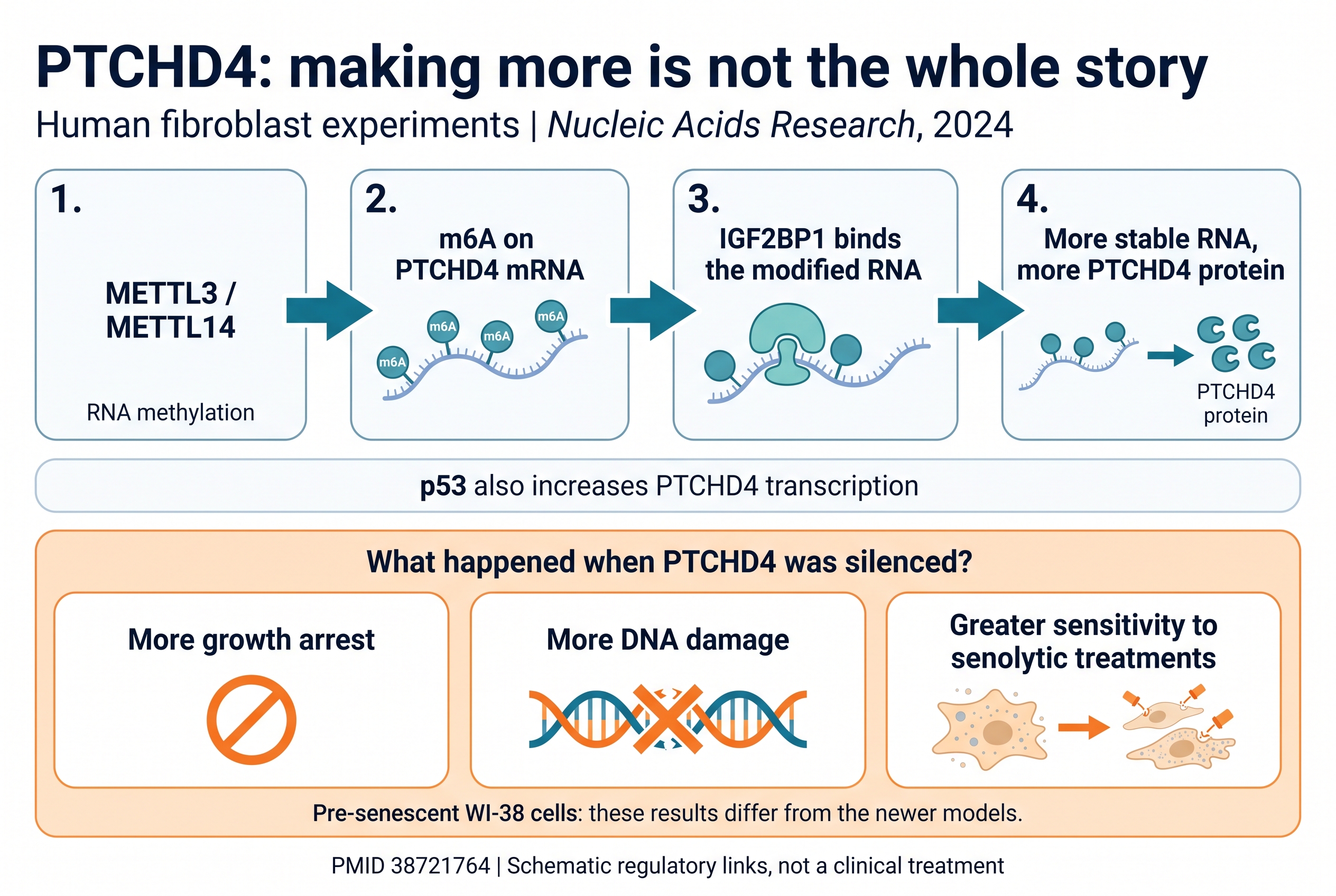
The 2024 Nucleic Acids Research study examined human fibroblasts, including cells approaching replicative senescence. Its investigators found a mechanism that helps cells produce more PTCHD4: the METTL3/METTL14 complex adds an RNA modification called N6-methyladenosine, or m6A, to PTCHD4 messenger RNA. IGF2BP1 recognizes the modified RNA and helps stabilize it. More stable RNA supports higher protein production. Nucleic Acids Research, 2024.
In that system, silencing PTCHD4 increased growth arrest and DNA damage. It also made pre-senescent cells more sensitive to the senolytic treatments tested in culture. These outcomes describe different aspects of cell behavior: becoming growth-arrested, accumulating damage, and surviving a subsequent challenge are not interchangeable endpoints.
The two papers therefore should not be merged into a single uninterrupted pathway. One reports attenuation of senescence in its deficiency models; the other reports increased arrest and damage after silencing in pre-senescent fibroblasts. Cell type, stage of senescence, experimental stress and the method of reducing PTCHD4 are possible explanations for the difference. That is a research hypothesis, not a reconciliation established by this article.
What can a molecular illustration honestly show?
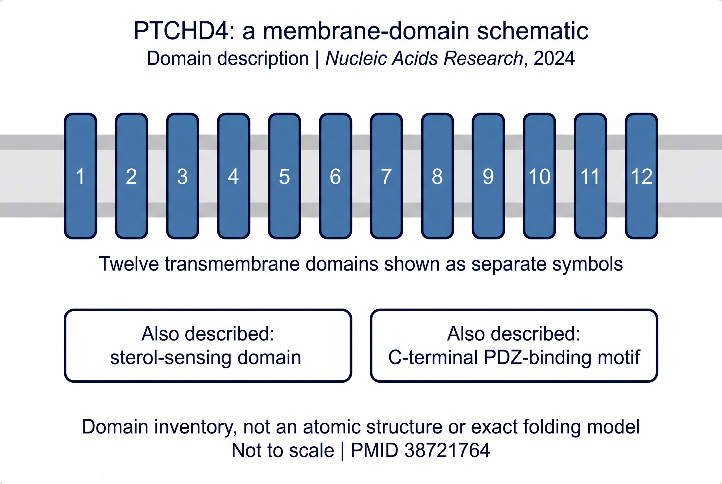
The earlier paper describes PTCHD4 as a member of the Patched protein family with 12 transmembrane domains, a sterol-sensing domain and a C-terminal motif capable of interacting with PDZ scaffold proteins. Its cell-fractionation experiments supported localization in membranous compartments rather than as a freely soluble cytosolic protein.
This supports a membrane-domain schematic. It does not justify presenting an AI-generated folded protein as an experimentally determined PTCHD4 structure. The illustration below deliberately omits atomic coordinates, residue-level binding sites and a proposed drug. Domain names also do not, by themselves, demonstrate a particular transport function or a direct PTCHD4–AKT interaction.
What would make this a stronger therapeutic lead?
The next useful question is whether a selective, timed intervention can reproduce the desired effects after disease has begun. Genetic deficiency and a medicine given to an adult are different interventions. Researchers would need to establish which cells to target, how much activity to change, and whether an apparent benefit comes with impaired function elsewhere.
Independent replication and a direct comparison of the differing cellular results would be especially valuable. For the lifespan claim, the full survival curves, animal characteristics, allocation methods, causes of death and adverse findings matter more than the word “extension” in an abstract. For lung disease, a promising model must eventually be followed by evidence in the relevant patients.
Neither paper establishes that a supplement, an AKT inhibitor or any available product safely reproduces a useful PTCHD4 intervention in people. There is no dosing guidance to extract from this coverage. The value of the finding is a more specific question for aging research, with a clear test of how that question might be wrong.
The membrane protein remains interesting. The different cells are the reason to keep looking.
Prepared by the Magellan Longevity Editorial Desk. AI-assisted research synthesis and original Nano Banana Pro illustrations. This is a focused comparison of two primary papers, not a systematic review. The new paper’s full-text limitations are stated above. No author endorsement is implied.
Educational, not medical advice.
The takeaway
PTCHD4 is a preclinical research lead whose effects differ across experimental contexts. The reported mouse and cell findings do not establish a safe human anti-aging intervention.
References
2 peer-reviewed sources, published 2024–2026, across 2 journals. Every citation links to its PubMed record.
- Aging Cell · 2026 · PMID 42725837 · DOI 10.1111/acel.70711
- Nucleic Acids Research · 2024 · PMID 38721764 · DOI 10.1093/nar/gkae322
Mechanisms and molecules in this article
Each links to its Magellan monograph — what it is, what it does, and the studies behind it.
Related reading
Rentosertib has renewed the debate over biological age. A randomized human experiment offers an important…
Tea, berries and spices were associated with a changing DNA-methylation clock in a small lifestyle trial.…
A 2009–2012 CDC survey analysis found periodontitis in 46% of dentate American adults 30 and older.…
Prefer the interactive version? Open this article inside the Magellan app →